![]() Single-Molecule
Biophysical Chemistry Group
Single-Molecule
Biophysical Chemistry Group
![]()
![]()
![]()
![]()
![]()
![]()
![]()
![]()
![]()
![]()
![]()
Nano-bioscience:
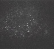
We are developing new single-molecule experiments that can be used to study protein-DNA interactions at nanometer precision levels. Current approaches to visualize enzymatic action of individual biomolecules rely on monitoring the molecular probes attached to individual enzyme molecules or their substrates. Successful probes include polystyrene beads and quantum dots in sub-micron size. Even more powerful probes in single-biomolecule detection are individual fluorophores in sub-nanometer size. We are also developing a single-molecule optical tweezers manipulation platform to apply calibrated piconewton forces to a single enzyme-DNA complex.
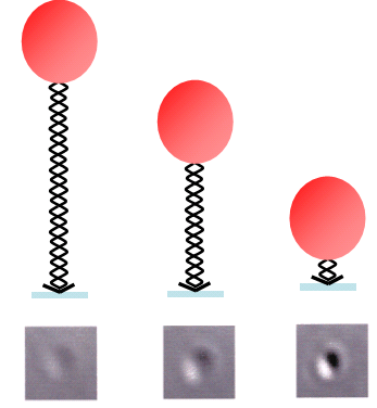
Even though the diffraction limit of optical microscopy is about 200 nm, an image spot can in fact be determined with nanometer precision through digital image processing. For example, the image of a 100 nm polystyrene bead is a two-dimensional spot that varies in intensity and spreads across an area contains a number of pixels in the digitized image. The intensity profile of this image spot can be fit into a Gaussian curve that is a function of position x and y in the image. The position of the fitted Gaussian image, and hence the centroid position of the bead, determines the positional resolution of the bead centroid to a nanometer precision. It is thus possible to image individual biomolecules with nanometer precision.
 |
 |
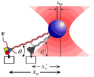 |
| (A). Single-Molecule Visualization through a sub-micron sized bead | (B). Single-Molecule Imaging through a single fluorophore | (C). Single-Molecule Manipulation through optical tweezers |
Last update: 2005.02.04 @ 12:15 PM